Tuesday, May 15, 2012

Discussion Questions from our Mentor

Histochemical Staining
1) What are the colour of the stained tissue?
     Purple Pink
2) What is the pattern of colour distribution in the stained tissue?
   Organelles are highlighted
3) Can you identify the nuclei of the cells? What colour are they?
    Dark purple
4) Can you identify the cytoplasm of the cells? What colour are they?
    Pink
5) What are the relative sizes and shapes of the cells in the tissue?
     Irregular
6) How many different types of cells do you think there are in the tissue?
     1
7) Are there any other observation you have identified? If so, describe them.
     The cells are irregular (aka. Adhesion is bad)


Immunohistochemical Staining
1) What are the colours in the tissue?
     Brown
2) What is the pattern of colour distribution in the stained tissue?
     It is around the cell membrane.
3) Are the colour and distribution similar to that in the first slide?
     No
4) The purpose was to determine the presence and location of a protein call vimentin. Which colour do you think represents vimentin? Why?
    Brown
5) Describe the location of vimentin in the tissue
     Cell membrane
6) What does the other colour represent?
    Other organelles
7) Are there any other observation you have identified? If so, describe them.
    Nope.


Fluorescence staining
1) Can you see the tissue using the fluorescence microscope? Does the tissue appear similar to the first and second slide?
   No, it is now azure color.
2) Describe what you observe. How many colours appear when using the fluoresence micropscope and what is the colour? 
   1, it is just blue.
3) Are there any other observation you had identified? If so, describe them.
     Not any special.

Our Achievements


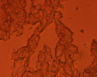
This is the pictures of our liver cancer cell after the histochemical staining! Eosin (the red dye) and hematoxylin (the blue dye) is added to the cell so thats why everything is redish purplish.


Problems we faced: Well our samples on the tissue decided to take a swim in the buffered solution. Some of the other team's samples literally went down the drain. We had to solve this by using a micropipette to put small droplets of PBS onto it, then taking it out with the micropipette again. We kept doing that until we thought it was clean of excess dye.


This is the picture of the liver cell under a fluorescence microscope and after it was stained with hoescht dye. The blue spots are the nucleus that has been stained and the other lighter blue is the the cytoplasm with the other organelles. Note that the organelles and the cytoplasm do not have any color. The blue is from the glowing nucleus.

Problems we faced: At first we couldn't see anything. No matter what the student mentors tried, we just saw one whole patch of black! We were advised to redo everything and then avoid placing the mounting media (a type of liquid that prevent air bubbles to be placed found under the coverslip). In the end, thanks to our student mentor, we got such brilliant pictures under the microscope!


This is Immunohistochemical staining! We first place a primary antibody that would seek and bind with the protein vinmentin. Vinmentin is a protein that is mostly found in cancerous cells, so we look for this in a potentially cancerous cell. Later we will put a secondary antibody, which would bind with the primary antibody and release an enzyme. Once we put a subtrate in, the enzyme and the substrate would form a brown precipitate that would appear at the areas the vinmentin is found.
Problems we faced: At first we couldn't really see anything, but later when we went to the microscope that has the camera and got this pictures. Otherwise everything quite smoothly


This is a picture of someone's hair :D


After all of this we had a presentation! 


Objective

-What does the group need to accomplish by the end of the project?
We should understand by then the use of the project in real life applications and also understand the reasons for using certain materials to do the analysis.

-How will you know whether or not your project has been successful?
When we use the UV light, we would find out if the project is sucessful

Overview

-What main theory or concept is the project based on?
The project is based on the cell anatomy and analysis of it.

-What do you already know about this theory or concept?
We know the basic of microscopes and the basic of cells anatomy.

Our team

Our team consist of Jun Wei, Charlene and Manish Muhuri!